Endoscopic Spine Surgery
Diagnostic Procedures
Pain Management Procedures
Traditional Spine Fusion Surgeries
Mini Spine Fusion and Spine Disc Replacement
Endoscopic Foraminotomy of the Lumbar and Cervical Spine
Endoscopic Foraminotomy is a minimally invasive spine surgery used to relieve pressure to spinal nerve roots, caused by compression from bone spurs, disc herniations, scar tissue, or excessive ligaments.
The purpose of the foraminotmy is to enarlge or open the narrowed neuro or spinal nerve root canal, so the nerves would have more room to move around without compression. Foramintomies are routine procedures in Atlantic Spine Center, offered to patients with arm pain (cervical foraminotomy) and leg pain (lumbar foraminotomy). After the procedure, usually the arm or leg pain and its associated tingling, numbness, and burning sensations disappear immediately.
multimedia
What are the Advantages of an Endoscopic Foraminotomy?
Endoscopic Foraminotomy surgery is a true minimally invasive spine surgery that include the following advantages:
- Minimally Invasive
- Short recovery
- High Success rate
- Local anesthesia
- Minimal or no blood loss
- Preservation of spinal mobility
- Small incision and Minimal scar tissue formation
- Same day surgery with no hospitalization (outpatient procedure)
What Conditions Can Endoscopic Foraminotomy Surgery Treat?
Lumbar Endoscopic Foraminotomy
- Arthritis of the spine / Bone spurs
- Bulging disc / Disc herniation
- Failed back surgery syndrome
- Foraminal narrowing (foraminal stenosis)
- Spine degeneration
- Radiculitis / Radiculopathy
- Sciatica
- Spinal slippage (spondylolisthesis)
- Spinal instability
- Spinal stenosis
Cervical Endoscopic Foraminotomy
- Cervical spine degenerative disease with spinal nerve compression
- Cervical spinal nerves pinched by disc herniations and bone spurs
How Is Endoscopic Foraminotomy Surgery Done?
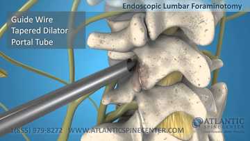
During endoscopic foraminotomy surgery, the patient is brought to the operative room. Under anesthesia, a small metal tube is inserted to the neuroforamen for direct visualization. The surgical tools are inserted through this tube so that your muscles do not need to be torn or cut open. The spinal nerve is found under direct visualization looking through the tube, and protected.
Under direct vision, bone spurs, scars, ligament overgrowth, protruded discs, and part of the troubled lumbar facet are removed with appropriate tools (eg, a laser, radiofrequency or mechanical tools) to enlarge the nerve hole and to release the compressed nerve(s). Finally, the tube is removed and the incision is closed with a stitch or two.
Upon completion, the patient is encouraged to walk around and is free to leave the surgical center, with a companion, the same day. After a follow-up visit with the surgeon the next day, the patient can go home for a quick recovery.

